Home
Uncategories
Hip Muscles Diagram Labeled / About the Muscular System : Related searches for muscles diagram labeled posterior view posterior view muscles diagramposterior muscle diagramposterior thigh muscles labeledhuman.
Hip Muscles Diagram Labeled / About the Muscular System : Related searches for muscles diagram labeled posterior view posterior view muscles diagramposterior muscle diagramposterior thigh muscles labeledhuman.
Hip Muscles Diagram Labeled / About the Muscular System : Related searches for muscles diagram labeled posterior view posterior view muscles diagramposterior muscle diagramposterior thigh muscles labeledhuman.. View the muscles of the upper and lower extremity in the diagrams below. Superficial muscles are the muscles closest to the skin surface and can usually be seen while a body is performing actions. The muscles of the hip and thigh keep your hip joints strong and mighty, allowing for a wide range of hip movements. It passes through the pelvis and extends to the thighbone, or femur. Forearm muscles anatomy, posterior arm muscles, muscles of the arm and forearm, forearm anatomy, arm muscles diagram, deep muscles of forearm, muscles in lower arm.
Smartdraw includes 1000s of professional healthcare and anatomy chart templates that you can modify and make your own. Everyone should list the structures within muscle. Learn vocabulary, terms and more with flashcards, games and other study tools. Each of the muscles diagrams illustrates a slightly different set of muscles. Now label the diagram in your workbook!
labeled muscles of lower leg - Yahoo Search Results ... from i.pinimg.com Forearm muscles anatomy, posterior arm muscles, muscles of the arm and forearm, forearm anatomy, arm muscles diagram, deep muscles of forearm, muscles in lower arm. Activity 4.6 labeled muscle diagram. Smartdraw includes 1000s of professional healthcare and anatomy chart templates that you can modify and make your own. Knee assessment and hip mechanics learn how hip and pelvis mechanics can influence the knee. This diagram depicts muscle labeled diagram. Label the major muscles of the body. The gastrocnemius has two parts or heads, which together create its diamond shape. The bones shown in the chest and hip region in the labeled human skeleton diagram are the ribs, vertebrae, pelvis, os coxae, sacrum and coccyx.
These muscles are separate in the abdomen, but they join together in the thigh.
Pubmed® comprises more than 30 million citations for biomedical literature from medline, life science journals, and online books. Most will label a diagram of muscle with its. Two individual muscles called the psoas major and the iliacus form the iliopsoas muscle. Human muscle system, the muscles of the human body that work the skeletal system, that are under voluntary control, and that are concerned with movement, posture, and balance. See more ideas about muscle diagram, medical anatomy, muscle anatomy. The adductor group is made up of: You should make a label that represents your brand and creativity. 6 photos of the diagram labelled of the hip muscles. Activity 4.6 labeled muscle diagram. Click on the labels below to find out more about your muscles. Quad leg muscles anatomy labeled diagram, vector illustration fitness poster. Now that you watched the video, you. Related searches for muscles diagram labeled posterior view posterior view muscles diagramposterior muscle diagramposterior thigh muscles labeledhuman.
Learn vocabulary, terms and more with flashcards, games and other study tools. Now label the diagram in your workbook! The adductor group is made up of: Superficial muscles are the muscles closest to the skin surface and can usually be seen while a body is performing actions. An easy and convenient way to make label is to generate some ideas first.
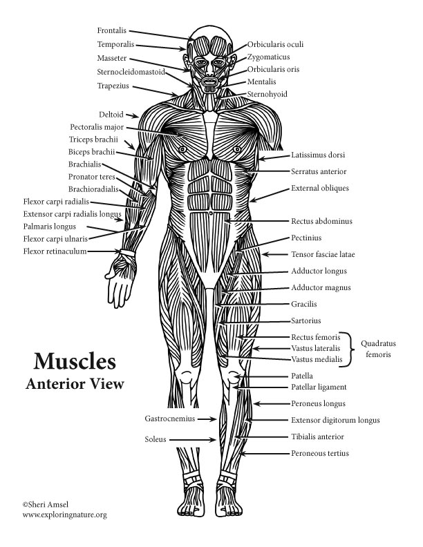
About the Muscular System from www.exploringnature.org The gastrocnemius is the larger calf muscle, forming the bulge visible beneath the skin. Extension and rotation of the hip origin: Two individual muscles called the psoas major and the iliacus form the iliopsoas muscle. Label the major muscles of the body. Sartorius is a unique muscle because it is the only knee flexor that originates anteriorly. If you're struggling, don't be hard on yourself. In human anatomy, the muscles of the hip joint are those muscles that cause movement in the hip. The bones shown in the chest and hip region in the labeled human skeleton diagram are the ribs, vertebrae, pelvis, os coxae, sacrum and coccyx.
Forearm muscles anatomy, posterior arm muscles, muscles of the arm and forearm, forearm anatomy, arm muscles diagram, deep muscles of forearm, muscles in lower arm.
The adductor group is made up of: Now that you watched the video, you. Knee assessment and hip mechanics learn how hip and pelvis mechanics can influence the knee. Press into the feet, lengthening the legs to press the hips up toward the ceiling. Feel the spine being pulled in opposite directions as you press the head. Label the major muscles of the body. An easy and convenient way to make label is to generate some ideas first. Everyone should list the structures within muscle. See more ideas about muscle diagram, medical anatomy, muscle anatomy. The bones shown in the chest and hip region in the labeled human skeleton diagram are the ribs, vertebrae, pelvis, os coxae, sacrum and coccyx. 6 photos of the diagram labelled of the hip muscles. These muscles are separate in the abdomen, but they join together in the thigh. Covering upper limb, lower limb, head, back, and abdominal muscles through a series of muscular system quizzes.
A complete list of muscular system quizzes; Smartdraw includes 1000s of professional healthcare and anatomy chart templates that you can modify and make your own. View the muscles of the upper and lower extremity in the diagrams below. Muscle and tendon anatomy of the hip (adductors, gluteal muscles (or buttocks), hamstring muscles, femoral muscle quadrices). Activity 4.6 labeled muscle diagram.
labeled muscles of lower leg - Yahoo Search Results | Leg ... from i.pinimg.com The bones shown in the chest and hip region in the labeled human skeleton diagram are the ribs, vertebrae, pelvis, os coxae, sacrum and coccyx. Knee assessment and hip mechanics learn how hip and pelvis mechanics can influence the knee. An easy and convenient way to make label is to generate some ideas first. Extension and rotation of the hip origin: Related searches for muscles diagram labeled posterior view posterior view muscles diagramposterior muscle diagramposterior thigh muscles labeledhuman. There are anterior muscles diagrams and posterior muscles diagrams. The calf muscle, on the back of the lower leg, is actually made up of two muscles: Knee assessment and hip mechanics online course:
There are anterior muscles diagrams and posterior muscles diagrams.
Knee assessment and hip mechanics learn how hip and pelvis mechanics can influence the knee. The adductor group is made up of: A basic human skeleton is studied in schools with a simple diagram. This diagram depicts muscle labeled diagram. Forearm muscles anatomy, posterior arm muscles, muscles of the arm and forearm, forearm anatomy, arm muscles diagram, deep muscles of forearm, muscles in lower arm. The calf muscle, on the back of the lower leg, is actually made up of two muscles: 6 photos of the diagram labelled of the hip muscles. Activity 4.6 labeled muscle diagram. Use the location, shape and surrounding structures to unlabeled diagram. Smartdraw includes 1000s of professional healthcare and anatomy chart templates that you can modify and make your own. Human muscle system, the muscles of the human body that work the skeletal system, that are under voluntary control, and that are concerned with movement, posture, and balance. From physical best activity guide: Quad leg muscles anatomy labeled diagram, vector illustration fitness poster.
The adductor group is made up of: hip muscles diagram. Superficial muscles are the muscles closest to the skin surface and can usually be seen while a body is performing actions.


0 Comments:
Posting Komentar